
 Write to WhatsApp
Author Roman Kartashov,
maxillofacial surgeon

Injuries to the skull may be accompanied by fractures of the bones forming the orbital cavity where the eyeball is located. If you or your child was injured, which led to "bruise" appearance under the eye, it is important to contact promptly a specialist who will determine whether there are any fractures. This is important, because orbital wall fracture may result in adverse consequences associated with visual function deterioration.
Orbital fracture symptoms
- Pain, swelling or bruising at the eye area
- Numbness of the cheeks and gums
- Nasal bleeding
- Eyes displacement, double vision
- Visual acuity decrease
- Facial deformation (facial skeleton fractures in extensive injuries).
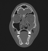
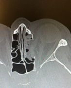
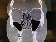
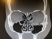
Orbital fracture diagnosis
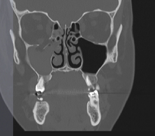
Computed tomography (CT) is the gold standard in the diagnosis of orbital fractures, allowing to accurately reproduce the look of the orbital skeleton and adjacent structures in several planes. 3D reconstruction gives accurate information on the number of bone fragments and their position.
Orbital fracture treatment
- Antibacterial therapy to prevent infection
- Symptomatic relief of pain, edema, subcutaneous hematoma
- Surgical reconstruction of skeleton, eyes position, draining of intraorbital hematomas
Orbital fractures may vary in both localization and severity. In general, it is important for the doctor to determine whether the surgery is required.
Without treatment, there is a possibility of adverse effects of post-traumatic deformity, blurred vision and eyes displacement. It is much more difficult to freat them than the "acute" injury.
Indication for surgery:
- Vision dysfunction (fracture often results in eye displacement, accompanied by double vision; growing hematoma may lead to optic nerve atrophy and vision loss; bone fragments may hinder the muscle contraction, which leads to restriction of eye movements);
- Facial skeleton deformation due to the bone fragments displacement, as seen by the changes in facial features, asymmetry, and disfigurement;
- Infraorbital nerve compression by displaced bone fragments, resulting in numbness of the cheek, a half of nose, lips and gums.
It is preferable to perform the surgery immediately after the fracture prior to the edema development. If swelling is already appeared, it is necessary to wait for 3-5 days.
Surgery options for orbital fracture
Very thin bones form orbital walls and it is impossible to return them to their previous position. However, there are several materials available for prosthesis: the patient's own calvarial bones, titan net and various synthetic prostheses.
As for orbital edge fractures, recovery of its shape is performed using the fixing titanium plates with screws, as the bone is thick enough here.
Rehabilitation after orbital fracture
After surgery, CT scan is performed to control the result. Both maxillofacial surgeon and ophthalmologist follow the patient.
As a rule, in uncomplicated postoperative period, swelling in the area of surgery begins to resolve in 3-4 days and only residual bruising in seen in 7-10 days.
Rehabilitation is aimed to restoration of visual function. Patient is advised to perform eye exercises, to avoid increased pressure in the nasal cavity during sneezing and blowing his nose.
Contraindications for surgery
- Severe traumatic brain injury
- Associated pathology in which any surgeries are contraindicated